- External URL
- Creation
-
Creator (Definite): Albert Frank Stanley KentDate: 1893
- Current Holder(s)
-
Description, references to Fig. 1:
'Sketch of the section of heart of adult Rat, showing junction of right auricle and ventricle. In the upper part of the figure is the outer wall of the right auricle, below and to the right is the wall of the right ventricle, and below and more to the left is the mass of tissue from which springs the auriculo-ventricular valve. The cavity of the heart is to the left. It is seen that a band of muscle runs almost completely round from auricle to ventricle, bordering closely upon the cavity of the heart. At the lower part this band becomes continuous with a network of branched muscle cells lying in the interval between auricle and ventricle. These branched fibres become continuous on the right with the muscular tissue of the ventricle.
Description, references to Fig. 2:
A small portion of Plate XII., Fig. 4, drawn under the one-twelfth oil immersion, to show the character of the muscular fibres lying in the fibrous tissue. They are seen to be sometimes fusiform, sometimes much branched cells, the branches being either comparatively thick or tapering off to very fine threads, which again become larger,. again branch, and are connected into a network with the branches of other similar cells. The nucleus usually causes a bulging of the celL Transverse striation is usually very well marked.


- No links match your filters. Clear Filters
-


-
 Cites
Cites Plate XII, Journal of Physiology 14 (4-5) (1893). Figs. 1-6 from A.F. Stanley Kent, 'Researches on the Structure and Function of the Mammalian Heart'.
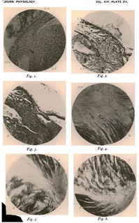
Description:'Explanation of Plate XII
Fig. 1. A photograph of a section of the heart of a young Rat, showing junction of left auricle anid left ventricle. The auriculo-ventricular valve is to the left. It is seen that the muscle fibres of the auiricle are continuous with those of the ventricle on the one hand, and with those of the valve on the other. Stained carminie.
Fig. 2. Photograph of section of the heart of aduilt Rat. Shows junction between left auricle and left ventricle. The lower mass of muiscular tissue is a part of the wall of the left ventricle, anid at the upper part of the figure and to the left is a part of the left auricle. There is no interruption of muscular continuity. Above and to the right is a blood-clot in the auricular cavity. Stained logwood.
Fig. 3. Photograph of a section of heart of adult Rat, showing junction between auricle and ventricle. The auriculo-ventricular valve is to the right of the figure.
Fig. 4. Photograph of fibrous tissue at auriculo-ventricular groove in heart of Monkey. Shows the branched and very slender muscular fibres imbedded in the fibrous tissue, and their connection with the fibres of the ventricle. To the left of the figure the network is fairly dense, on the right the fibres are somewhat thicker. At the lower part of the figure the modeof termination of the ventricular fibres is well seen. A process-block of a drawing of this section has been printed with the text.
Fig. 5. Photograph of auriculo-ventricular groove in heart of Monkey. This figure shows the way in which the auricular fibres branch on reaching the fibrous tissue. At the lower part of the figure on the right a stellate mass of auricular muscle is seen, some of the fibres of which become continuous with some of the scattered branched muscle cells lying in the fibrous tissue. At the top of the figure the tapering terminations of the ventricular bundles are seen, and on the right a few of the branched fibres lying in the fibrous tissue.
Fig. 6. Photograph of auriculo-ventricular groove in heart of Monkey. Shows the way in which the auricular muscular tissue becomes broken up into stellate masses as it approaches the fibrous tissue, and the manner in which these stellate masses become connected with one another by lateral branches. To the right of the figure a few branched cells are seen.' (253-254)
Fig. 1 in the article:
'Fig. 1, Plate XII., represents part of a coronal section of the heart of a newly-born rat, the point of junction of the auricle and ventricle having been photographed. It is seen here that the cells forming the mass of the ventricular wail are somewhat different fronm the usual form of cardiac muscular fibres, being more fusiform and having more sharply tapering ends. The nuclei also have a more or less pronounced fusiform outline and strongly resemble the nuclei of non-striped muscle cells. At the upper part of the ventricle and in the angle formied by the auricular wall the muscular fibres of the ventricle are seen to have a direction somewhat different to that taken by the auricular fibres, but at about the centre of the isthmus the auricular fibres are seen to sweep freely down into the substance of the ventricular wall and other fibres are to be observed coursing from the auricle into the basal part of the valve. At this stage therefore it would appear that a muscular continuity exists between the auricle and ventricle, and also between the auricle and base of the auriculo-ventricular valve.' (240)
Plate XII as a whole in the article:
'But in addition to this comparatively simple [muscular fibre] mode of connection [between auricles and ventricles], we have in the Monkey remarkably well developed a second and far more complicated system of communicating fibres, indications of whose presence we may find lower down in the scale, inasmuch as they are recognisable even in the rat...
In the Monkey these fibres are far more perfectly developed and present a conmplete network permeating the fibrous connective tissue and extending through from auricle to ventricle. Not only are these fibres present in the connective tissue but the normal cardiac muscle on approaching the groove appears to split up into similar fibres and becomes connected with the network of cells previously mentioned, and in this way a second system of communicating fibres is established between auricle and ventricle (see figures, Plate XII.).' (243)
