- Tags
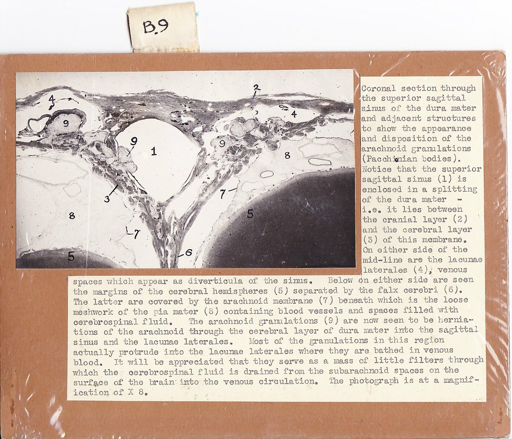
Coronal section through the superior sagittal sinus of the dura mater and adjacent structures to show the appearance and disposition of the arachnoid granulations (Pacchionian bodies). Notice that the superior sagittal sinus (1) is enclosed in a splitting of the dura mater – i.e. it lies between the cranial layer (2) and the cerebral layer (3) of this membrane. On either side of the mid-line are the lacunae laterales (4), venous spaces which appear as diverticula of the sinus. Below on either side are seen the margins of the cerebral hemispheres (5) separated by the falx cerebri (6). The latter are covered by the arachnoid membrane (7) beneath which is the loose meshwork of the pia mater (8) containing blood vessels and spaces filled with cerebrospinal fluid. The arachnoid granulations (9) are now seen to be herniations of the arachnoid through the cerebral layer of the dura mater into the sagittal sinus and the lacunae laterales. Most of the granulations in this region actually protrude into the lacunae laterales where they are bathed in venous blood. It will be appreciated that they serve as a mass of little filters through which the cerebrospinal fluid is drained from the subarachnoid spaces on the surface of the brain into the venous circulation. The photograph is at magnification of X 8.
Original card